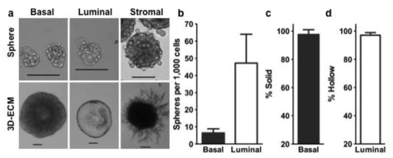
Figure 1.

Characteristics of mammospheres derived from different fractions of fluorescence-activated cell sorting (FACS) sorted cells from wild type C57BL/6 mice (a). Single spheres were further plated in Matrigel to form corresponding morphologically unique 3-dimentional extracellular matrix (3D-ECM) structures (a). Bar graphs showing the sphere formation efficiency (b) and percentage of solid (c) or hollow (d) structures in 3D-ECM culture. Scale bars, 100 μm. The mean and standard deviation values, and the number of replicates used for each plotted figures are stated in the Results.
