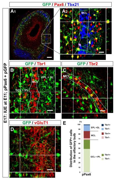
Figure 6. Impaired mitral cell differentiation by sustained Pax6 expression in embryonic olfactory bulb.
(A – D) Horizontal sections of E17 OB electroporated with pPax6 and pGFP. GFP+ cells (Cy2, green) are Pax6+ (A; Alexa 555, red), and localize both inside and outside the mitral cell layer that is defined with Tbx21 (A; Alexa 647, blue). GFP+ cells positioned inside the mitral cell layer express Tbr1 (B; Alexa 555, red), Tbr2 (C; Alexa 555, red), and vGluT1 (D; Alexa 555, red). In contrast, GFP+ cells found outside the mitral cell layer are mostly negative for these molecules. (E) Graph showing distribution and Tbr1 expression of GFP+ cells in E17 OB electroporated with pPax6 and pGFP. The graph was made in the same way as described in Figure 3J. Data acquired from 4 OBs were averaged. Scale bars, 50 μm.

