FIGURE 2.

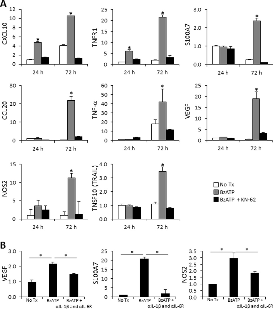
Cutaneous proinflammatory innate immune responses induced ex vivo following signaling through the P2X7R. (A) Bar graphs demonstrate the relative fold change in mRNA expression of CXCL10, TNFR1, S100A7, CCL20, TNF-α, VEGF, NOS2, and TRAIL 24 and 72 h following cutaneous injections with 350 µM of BzATP ± 1 µM KN-62 normalized to 24 h PBS injected controls (No Tx). (B) Examination of VEGF, S100A7, and NOS2 mRNA fold-change, normalized to PBS control (No Tx), 72 h following treatment with BzATP ± IL-1β and IL-6Rα antibodies. Fold-change was determined using the relative qRT-PCR 2−ΔΔCt method. Data expressed as mean ± SD of triplicates. Asterisk indicates a significant difference compared to no Tx for each time point, unless otherwise indicated, p < 0.05. Representative of three independent experiments.
