
FIGURE 3.
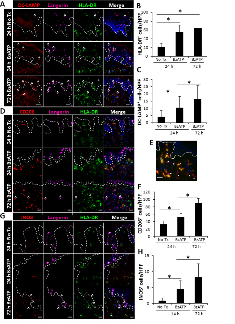
Increased expression of inflammatory markers on cutaneous DCs following ex vivo stimulation with BzATP. Skin explants were injected with 350 µM of BzATP or PBS (No Tx). 24 and 72 h following injections skin sections were immunofluorescently labeled with Langerin, HLA-DR, and (A) DC-LAMP, (D) CD206, or (G) iNOS. Dotted line represents epidermal-dermal junction and ^ indicates representative Langerin+ cells that are expressing DC-LAMP, CD206, or iNOS. Merged panels include all three stains and DAPI nuclear counter-stain. (E) Characterization of CD206+ cells 72 h following BzATP treatment. CD206-red and CD163-green. * indicates representative DCs (CD206+CD163−) and ^ indicates representative macrophages (CD206+CD163+). 40× original magnification, bar= 20µm. (B, C, F, H) Quantitation of cells positive for HLA-DR, DC-LAMP, CD206, or iNOS. Bars indicate the mean ± SD of 11–15 replicates. Asterisk indicates a significant difference, p< 0.05.