
FIGURE 4.
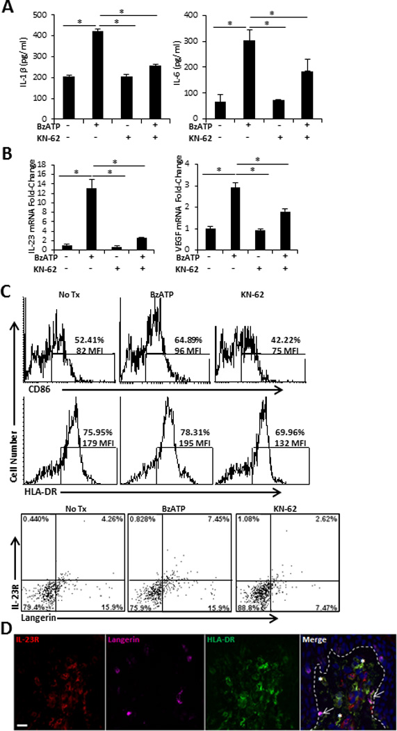
Direct stimulation of human smiDCs with P2X7R agonist. SmiDCs were cultured in the presence of the indicated treatments for 24 h and analyzed by (A) ELISA for the detection of secreted IL-1β and IL-6 in the culture supernatants and (B) qRT-PCR for the fold-change of IL-23 and VEGF expression. Data are representative of three-eight independent experiments; bars represent the means ± SD from triplicates. Asterisks indicate a significant difference between indicated groups, p < 0.05. (C) SmiDCs were stained with HLA-DR-, CD86-, IL-23R-, and Langerin-Abs. Top histograms are CD86 and HLA-DR expression; numbers represent percent positive and MFI within the gated region. Bottom dot-plots signify Langerin and IL-23R expression; numbers represent the percent positive within the respective quadrant. Flow cytometry data is one representative of four independent subjects with similar results. (D) Cutaneous explants were injected with 350 µM of BzATP and immunofluorescently labeled with IL-23R, Langerin, and HLA-DR 72 hr following treatment. Dashed line indicates epidermal-dermal junction. 40× original magnification, bar= 20µm. Data is one representative of two independent experiments.