
FIGURE 7.
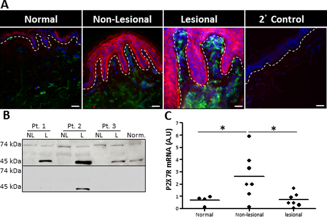
Lesional and non-lesional psoriatic skin exhibit enhanced P2X7R expression. (A) Human healthy donor and psoriatic non-lesional and lesional skin explants were immunofluorescently labeled with markers for P2X7R (red) and HLA-DR (green), plus DAPI (blue) nuclear counter-stain. As a staining control normal skin was stained with the secondary anti-rabbit-Cy3 Abalone (2° Control). Original magnification is 40×, bar = 20µm. Dotted line indicates the epidermal-dermal junction. Representative of four independent experiments. (B) Protein extracts were isolated from normal healthy skin and non-lesional and lesional psoriatic skin. Western blot analysis demonstrated the presence of two specific P2X7R variants, a 74 kDa and 45 kDa protein (upper panel). P2X7R specificity was confirmed with a blocking peptide (lower panel). Three individual patients are presented, which is representative of seven individual donors examined. (C) Scatter plot demonstrates arbitrary units of P2X7R mRNA expression normalized to B2M utilizing the 2−ΔCt method. Each dot represents an individual biopsy from normal, psoriatic non-lesional, or psoriatic lesional skin. Asterisk indicates a significant difference between indicated groups, p < 0.05.