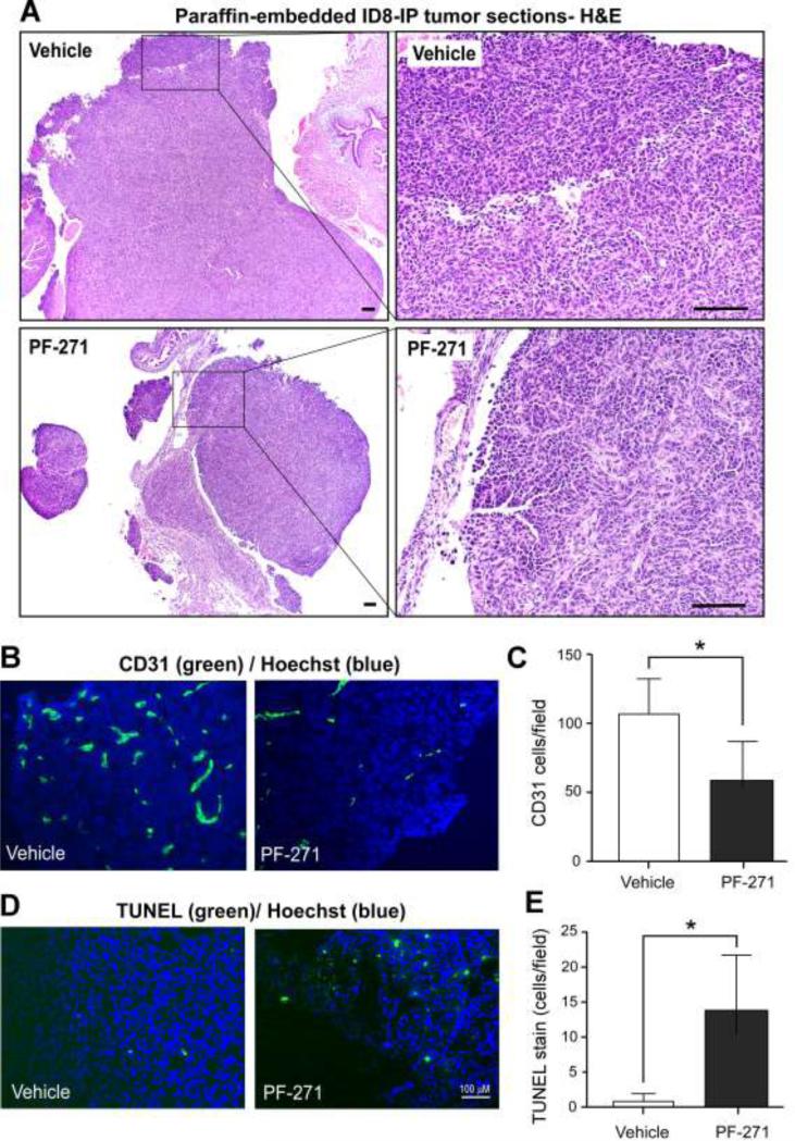
Figure 7.
Alterations in ID8-IP tumor-associated endothelial cells and tumor apoptosis in PF-271-treated mice. (A) Representative x40 images of paraffin-embedded H&E stained tumors from vehicle- or PF-271-treated mice. Right, boxed region x40 image at x200 magnification. Scale is 100 μM. (B) Representative fluorescent microscopy images of anti-CD31 antibody (green) and cell nuclei (Hoechst, blue) staining within ID8-IP tumors from vehicle- or PF-271-treated mice. (C) Fluorescent images from five random fields (at 10X) from 3 different ID8-IP tumors from vehicle- or PF-271-treated mice were acquired and the number of CD31-positive cells per field were enumerated using Image J. Values are means (+/- SD) (*p<0.05). (D) Representative fluorescent microscopy images of TUNEL (green) and cell nuclei (Hoechst, blue) staining within ID8-IP tumors from vehicle- or PF-271-treated mice. Scale is 100 μM. (E) Images were acquired and enumerated as above. Values are means (+/- SD) (*p<0.05).

