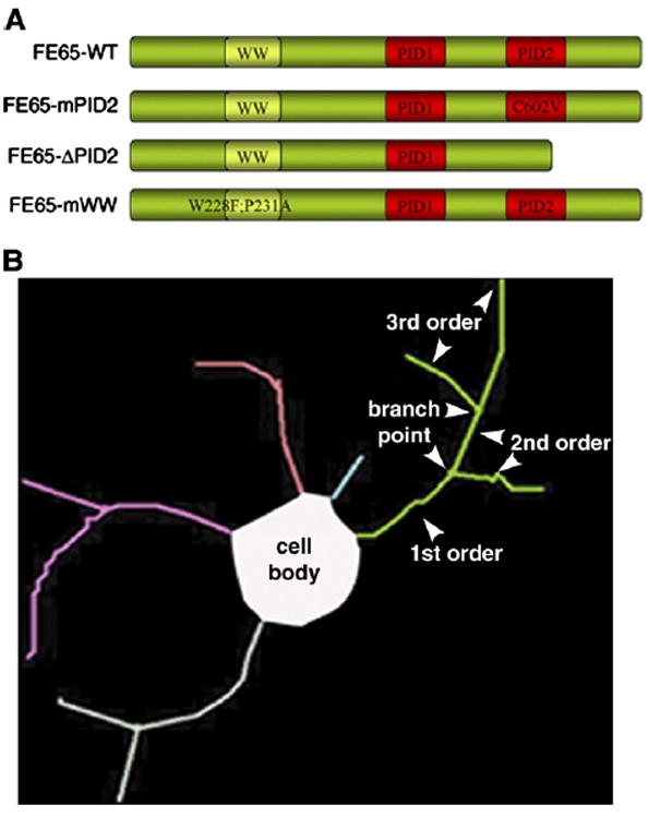
Fig. 1.

Scheme of viruses and tracing method used. (A) Schematic of the FE65 dominant-negative viral constructs used. FE65-ΔPID2 lacks the entire PID2 domain. FE65-mPID2 and FE65-mWW contain the indicated point mutations, which disrupt the interaction of FE65 with APP and Mena, respectively. (B) Example of a Neurolucida trace depicting the terms used (i.e. neurite order, branch points, etc.).
