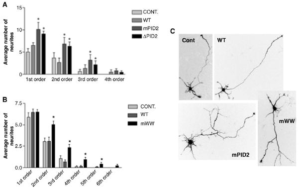
Fig. 2.

FE65 negatively regulates neurite branching as part of a macromolecular complex. (A, B) Expression of the FE65 PID2 (A) and WW (B) mutants illustrated in Fig. 1A increase neurite branching. Neurons were infected with adenovirus expressing wild type FE65 (WT), FE65-ΔPID2 (ΔPID2), FE65-mPID2 (mPID2) or GFP virus (CONT) 3h after plating. Neurites of infected 1 DIV (A) or 2 DIV (B) neurons were traced, counted and plotted as a function of neurite order number. 1st order neurites are defined as the segments extending directly from the cell soma. Branches correspond to 2nd order and higher neurites (see illustration in Fig. 1B). Branches correspond to 2nd order and higher neurites (n=30 neurons per treatment; *p<0.05). The data shown represent the mean ± S.E.M. (C) Examples of hippocampal neurons expressing GFP (Cont), wild type FE65 (WT), FE65-mPID2 (mPID2) and FE65-mWW (mWW). Scale bar equals 10 μm.
