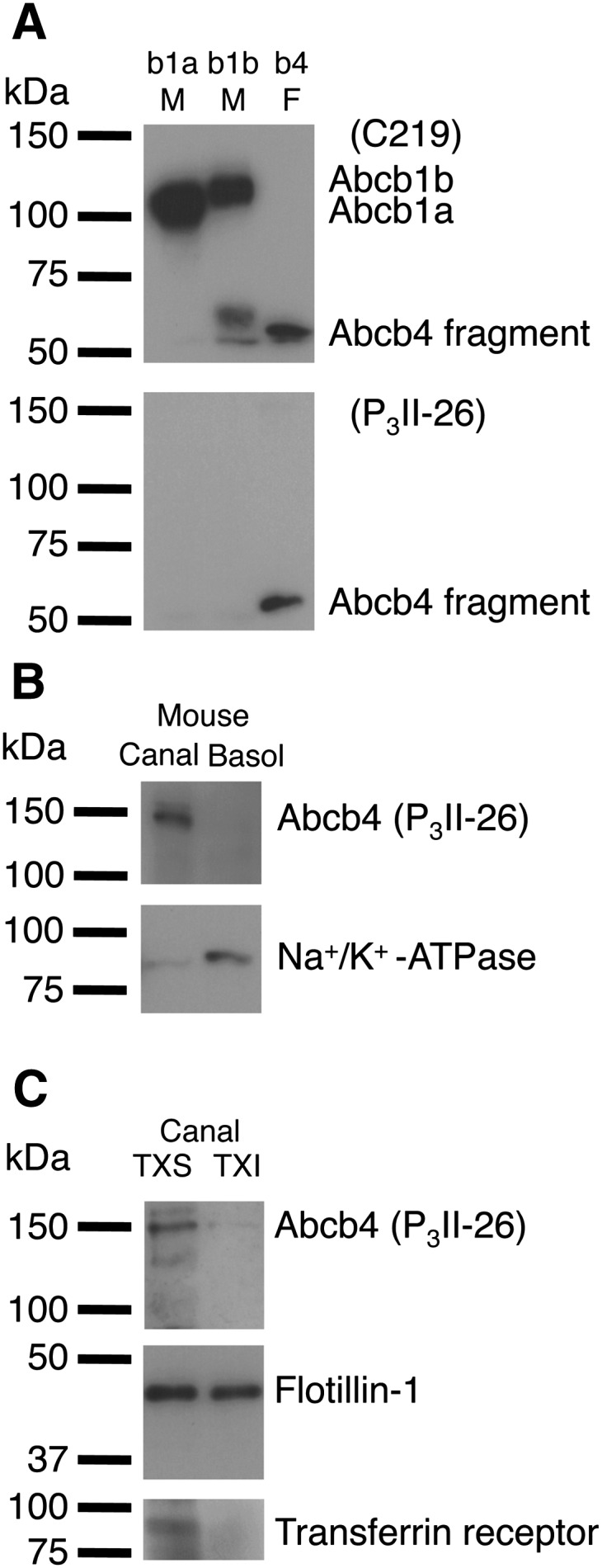
Fig. 1.

Distribution of Abcb4 between canalicular nonraft and raft membranes in mouse hepatocytes. A: Immunoreactivity of mouse Abcb1a, Abcb1b, and Abcb4 with C219 and P3II-26 monoclonal antibodies. Mouse Abcb1a membranes (b1a M) (2 μg of protein), mouse Abcb1b membranes (b1b M) (2 μg of protein), and mouse Abcb4 recombinant fragment (amino acids 352–708) (b4 F) (27.2 ng of protein) were separated by 10% SDS/PAGE. B: Mouse canalicular liver plasma membranes (Canal) and basolateral liver plasma membranes (Basol) (37.4 μg of protein) were separated by 10% SDS/PAGE. C: TXS and TXI fractions of mouse canalicular liver plasma membranes (equal volumes) were separated by 10% or 12% SDS/PAGE. Abcb4, Na+/K+-ATPase (basolateral marker), flotillin-1 (raft marker), and transferrin receptor (nonraft marker) were detected with specific antibodies.
