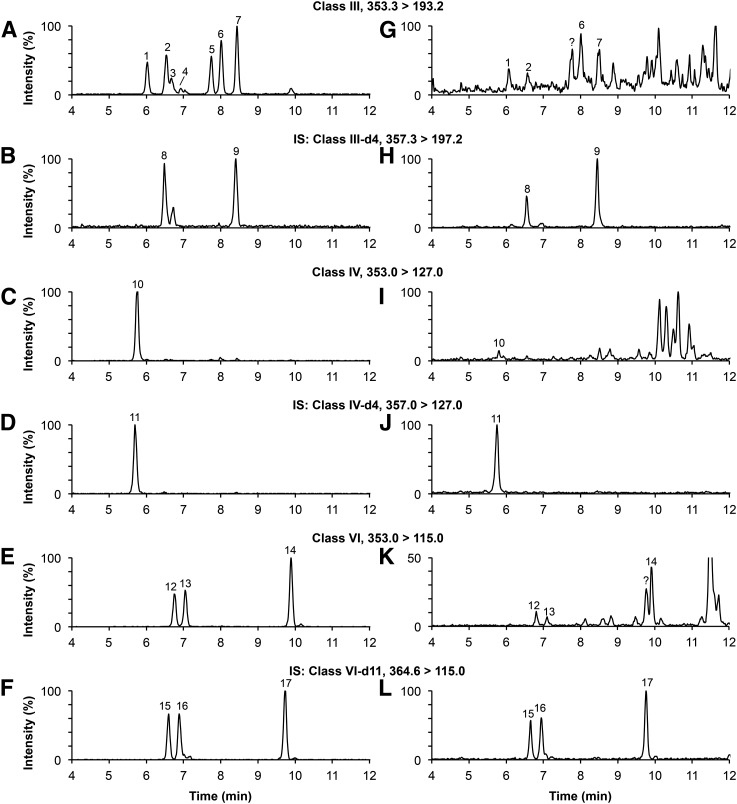
Fig. 2.
Mass chromatograms of a standard solution (A–F) and of a typical plasma sample spiked with 50 pg of each internal standard (G–L). Peak identification (see Fig. 1 for structure): 1, 8-iso-15(R)-PGF2α; 2, 8-iso-PGF2α; 3, 8-iso-PGF2β; 4, 11β-PGF2α; 5, 15(R)-PGF2α; 6, 5-trans-PGF2α; 7, PGF2α; 8, 8-iso-PGF2α-d4; 9, PGF2α-d4; 10, iPF2α-IV; 11, iPF2α-IV-d4; 12, iPF2α-VI; 13, 5-iPF2α-VI; 14, (±)5-8,12-iso-iPF2α-VI; 15, iPF2α-VI-d11; 16, 5-iPF2α-VI-d11; 17, (±)5-8,12-iso-iPF2α-VI-d11; ?, unknown compounds. IS, deuterated internal standards.

