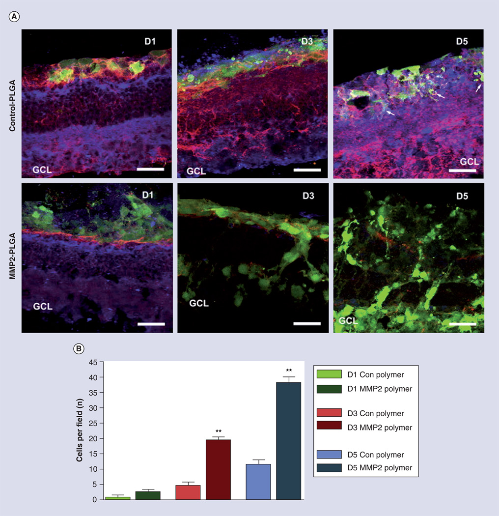
Figure 4. The controlled delivery of active MMP2 from electrospun fibrous scaffold enhances green fluorescent protein and retinal progenitors cells migration into degenerated Rho−/− retinal explants by degrading inhibitory matrix molecules CD44 and neurocan.
(A) Retinal explants were cultured in the absence (control PLGA) or presence (MMP2–PLGA) of active MMP2 for 1, 3 and 5 days at 37°C, and subsequently fixed, cryosectioned and immunolabeled for green fluorescent protein (green), CD44 (red) and neurocan (blue). (B) The average number of cells that cross from the polymer into the retinal explant per microscopic field.
(A) Magnification 40×, scale bar = 25 µm.
*p < 0.05; **p < 0.001.
GCL: Ganglion cell layer; PLGA: Polylactic-co-glycolic acid.
Reproduced with permission from [42]. © Elsevier (2009).
Color figure can be found online at www.expert-reviews.com/doi/suppl/10.1586/eop.12.56

