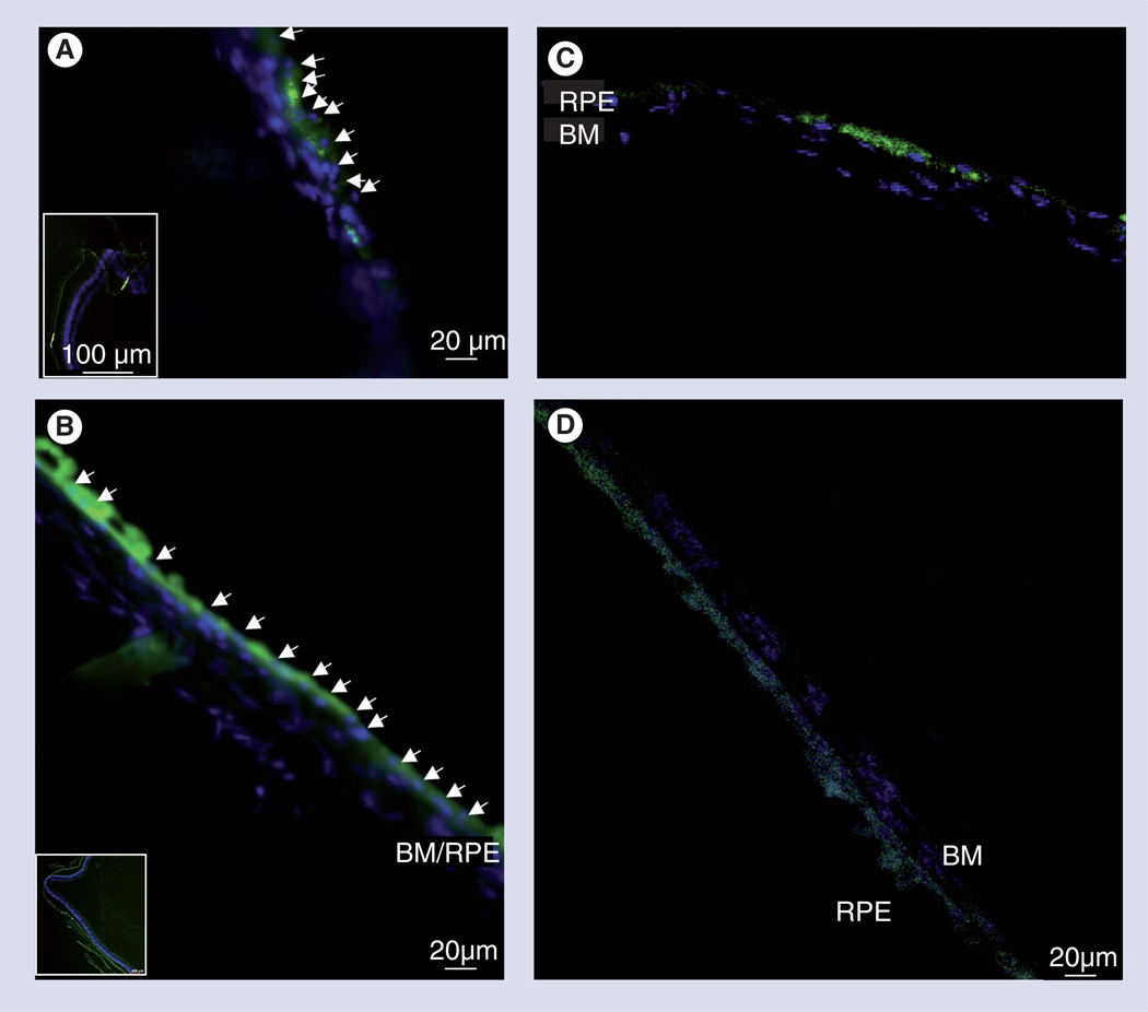
Figure 6. In vivo adult subretinal transplantation of green fluorescent protein and retinal progenitors cells in haluronate and methylcellulose self-aggregating hydrogel, assayed at 4 weeks post-transplantation.
(A) Control transplantation in saline vehicle show non-contiguous cellular integration and localized cellular groupings (inset) atop BM, suggestive of cellular aggregation pre- or post-transplantation. (B) Transplantation in haluronate and methylcellulose (HAMC) shows contiguous areas of RPE integration over large areas of retina (inset), suggesting HAMC maintains cellular distribution during injection and preventing aggregation pre- or post-transplantation. Arrowheads indicate location of nuclei of transplanted cells. Confocal images of cuboidal RPE cells sitting atop Bruch’s membrane after injection in (C) saline and (D) HAMC (Hoechst and green fluorescent protein).
BM: Bruch’s membrane; RPE: Retinal pigment epithelium.
Adapted with permission from [50]. © Elsevier 2010.
Color figure can be found online at www.expert-reviews.com/doi/suppl/10.1586/eop.12.56

