Table 1.
Polymers used in the scaffold production for the transplantation of retinal progenitors cells and their properties.
| Polymer | Chemical structure | Scaffold type |
Mechanical stiffness (modulus) |
Biodegradation | Notes |
|---|---|---|---|---|---|
| PLA | 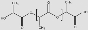 |
Cylinder | 2.8–4.1 GPa (l) 1.4–2.8 GPa (d,l) [73] |
12–24 months (depends on crystallinity) [15] | Modulus and degradation depends on crystallinity |
| PLGA |  |
Cylinder or fiber | 1.4–2.8 Gpa [73] | 1–6 months [15] | Modulus and degradation depends on lactide to glycolide ratio |
| PCL |  |
Cylinder or fiber | 206–344 Mpa [73] | >24 months [15] | |
| PGS | 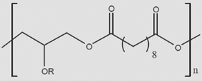 |
Cylinder | 0.282 ± 0.025 Mpa [74] | 1–2 months [74] | |
| PMMA | 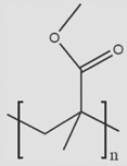 |
Cylinder | 1.8–3.1 Gpa [75] | Nondegradable | |
| Chitosan | 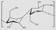 |
Fiber | 159.4 ± 40.0 Mpa (nanofibers) [76] | Enzymatically by chitosanase and lysozyme | Rate of degradation is dependent on primary nitrogen substitutions |
| HAMC | 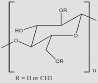 |
Hydrogel | 1–100 Pa [49] | 5–15 days [49] | Modulus and degradation rate are dependent on ratio of polymers |
| PNIPAAm | 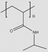 |
Hydrogel | N/A | N/A | Grafted to collagen to create self-aggregating hydrogel |
HAMC: Hyaluronan/methyl cellulose; N/A: Not applicable; PCL: Polycaprolactone; PGS: Polyglycerol sebacate; PLA: Polylactic acid; PLGA: Polylactic-co-glycolic acid; PMMA: Polymethyl methacrylate; PNIPAAm: Poly(N-isopropylacryl amide).
