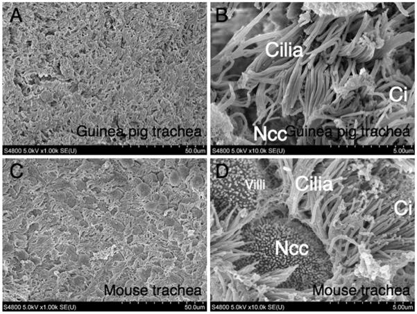
Fig. 1.
Scanning electron microscopy (SEM). Tracheal epithelia from (A and B) guinea pig and (C and D) mouse were evaluated by SEM. More abundant non-ciliated column epithelial cells were observed in the mouse epithelia (C and D) as compared with guinea pigs (A and B). Ci, ciliated cell; Ncc, non-ciliated epithelial cell. Scale bars in A and C = 50 μm, B and D = 5 μm.

