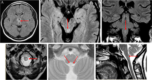
Figure 1.

Typical brain MRI lesions in neuromyelitis optica. Representative MRI of six NMO patients; Upper row: FLAIR; lower row: T2W, A. Lesions in hypothalamic region, B. Lesions in periaqueductal matter in mesencephalon, C and D. Lesions in medulla oblongata, E and F. Lesions in area postrema of the medulla oblongata.
