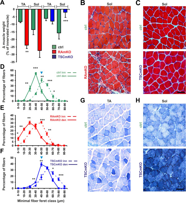
Figure 5.
Muscle atrophy induced by denervation. (A) Loss (Δ) of muscle weight in tibialis anterior (TA) and soleus (Sol) muscles after six days of denervation using mice of the indicated genotype. Data are expressed as percentage of weight loss compared to the non-denervated contralateral muscle in the same mouse. N ≥4 mice for RAmKO and control littermates (ctrl); N ≥5 mice for TSCmKO and control littermates. (B, C) H&E staining of soleus muscle after six days of denervation in mice of the indicated genotype. (D-F) Fiber size distribution in soleus muscle after six days of denervation (solid line) and in the contralateral, non-denervated muscle (dashed line) of mice with the indicated genotype. Note that the most frequent fiber size in the denervated TSCmKO muscle is the same as that of innervated control muscle (blue arrowheads). N ≥4 for RAmKO and control littermates; N = 5 for TSCmKO and control littermates. (G, H) NADH-TR staining of TA and soleus muscles after six days of denervation in control and TSCmKO mice. Scale bars (B, C, G, H) = 50 μm. Quantification represent mean ± SEM. P-values are ***P <0.001; **P <0.01; *P <0.05 using the Student’s t-test.

