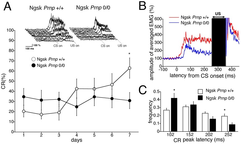
Figure 3. Impaired eyeblink conditioning in 40-week-old Ngsk Prnp 0/0 mice.
(A) CR development during a 7-day acquisition session in control (open circle, n = 10) and Ngsk Prnp 0/0 (experimental mice; closed circle, n = 10) mice aged 40 weeks. The top inset shows individual response topographies of averaged EMGs until US onset in control and experimental mice on day 7. (B) Averaged EMG amplitudes during the acquisition session on day 7. All EMG amplitudes obtained in 1 day were summed, representing the average of the eyelid responses. (C) Frequency histogram showing the peak CR latencies. The CR on day 7 was binned into time windows of 52–102, 102–152, 152–202, and 202–252 ms measured from CS onset in experimental mice (closed column) and control mice (open column). Data points represent the mean ± SEM. *p<0.05.

