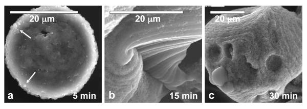
Figure 5.

Evolution of the calcium phosphate spherules formed at high concentration of PD (50 μg/mL) at different reactions times: (a) 5 min, (b) 15 min and (c) 30 min. Arrows in (a) point to the ACP precursor droplets that coalesced into a larger structure. A roughening of the texture within the molten spherulite was observed over time.
