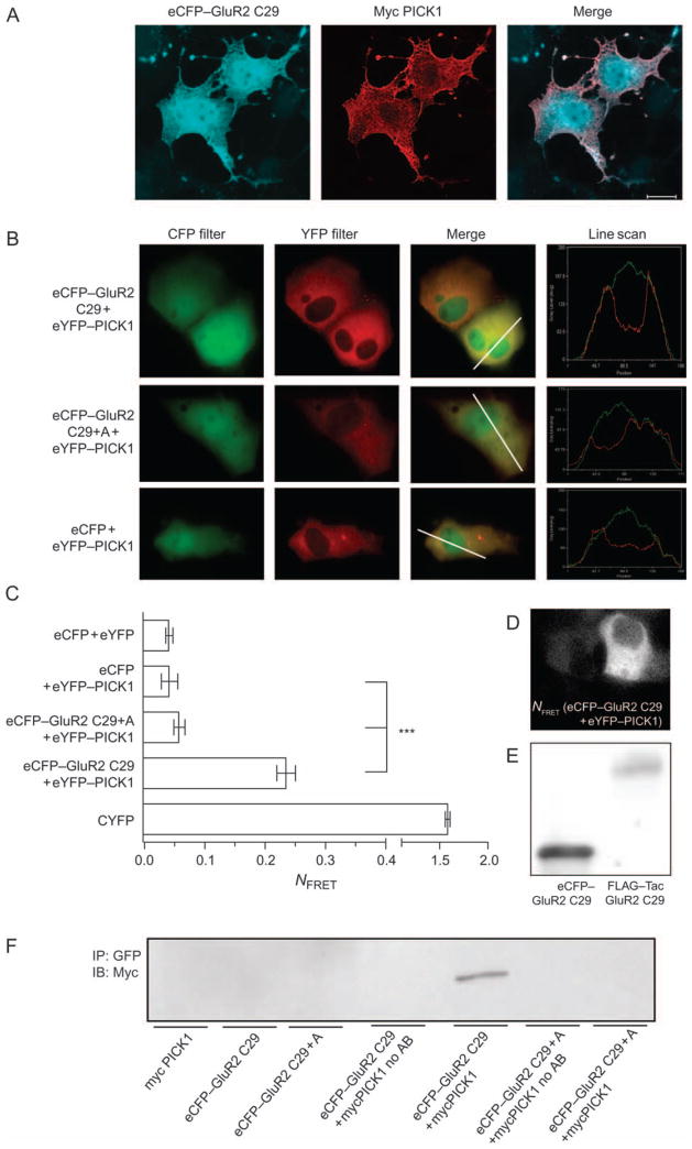
Figure 7. The interaction of PICK1 with a cytosolic ligand does not cause clustering of PICK1.
A) COS7 cells transiently coexpressing eCPF–GluR2 C29 (eCFP with the 29 C-terminal residues of GluR2 fused to the C-terminus) and mycPICK1. Cells were fixed, permeabilized and stained with anti-Myc. eCFP was visualized by CFP fluorescence. eCFP–GluR2 C29 is shown in blue (left column) and mycPICK1 in red (middle column). Right column shows the merged pictures. White bar = 10 μm. The data shown are representative of three experiments. B) COS7 cells expressing eCPF–GluR2 C29 and eYFP–PICK1, eCPF–GluR2 C29 + Ala (non-binding control) and eYFP–PICK1 or eCFP and eYFP–PICK1. The first and second columns from the left show the images obtained with CFP and YFP filter sets, while the third column represents a merged image of CFP and YFP. The fourth column shows line scan histograms illustrating cellular colocalization. The images shown are representative of three independent experiments. C) Normalized FRET efficiency is given for eCFP–GluR2 C29 and eYFP–PICK1 (n = 29), eCFP–GluR2 C29 + Ala and YFP–PICK1 (n = 28) and eCFP cotransfected with YFP–PICK1 (n = 22); as controls, we used CYFP (a covalent fusion of CFP and YFP) (n = 22) and cotransfection of CFP and YFP vectors (n = 20). All bars represent data from three experimental days (mean ± SEM). Statistical analysis was done using ANOVA, post hoc Bonferroni’s test for multiple comparisons (***p < 0.001). D) Distribution of the FRET signal corrected for bleed-through in COS7 cells expressing eCFP–GluR2 C29 and eYFP–PICK1. E) Western blot of eCFP–GluR2 C29 and FLAG–TacGluR2 C29 transfected in parallel. The two proteins were visualized with an antibody against the C-terminal 20 residues of GluR2 (Santa Cruz). F) Coimmunoprecipitation of myc-tagged PICK1 with eCFP–GluR2 C29 but not with eCFP–GluR2 C29 + Ala in agreement with the FRET measurements and a significant interaction. The experiments were carried out using lysates from COS-7 cells transiently expressing mycPICK1, eCFP–GluR2 C29, eCFP–GluR2 C29 + Ala, eCFP–GluR2 C29 together with mycPICK1 or eCFP–GluR2 C29 + Ala together with mycPICK1. The immunoprecipitations were done with a mouse monoclonal anti-green fluorescent protein antibody and immunoblotting with a rabbit anti-myc antibody. No antibody refers to no antibody in the immunoprecipitate.

