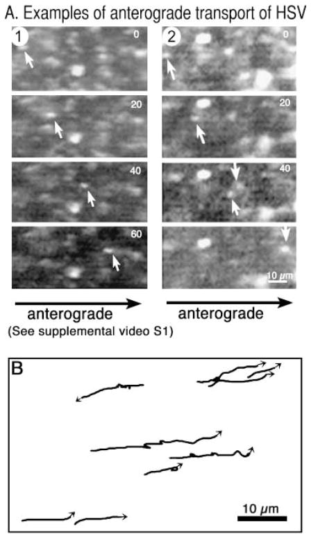
Fig. 2.
Fast anterograde transport of GFP-HSV in the squid giant axon. (A) Video sequence (39 frames, 2.67-s time lapse) of an individual VP16-GFP labelled particle travelling in the anterograde direction after injection into the axon. Adjacent stationary particles testify to the specificity of this movement. Some quenching of the GFP occurs as a result of the large amount of laser power required to obtain images rapidly of these small moving particles. (Please refer to Supplementary Material section for information on how to view the video clip in full.) (B) Tracings of VP16-GFP particle movements from a 50-frame/138-s sequence showing that viral particles may pause up to three times during this time span, some displaying short retrograde movements during pauses. In this field, one particle travels retrograde.

