Fig. 3.

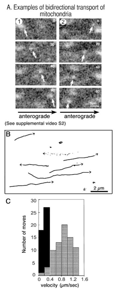
Mitochondria travel more slowly than HSV and in both directions. (A) Mitochondria, stained with the cell-permeable mitochondrial dye, R123, and imaged in the same axon as Fig. 2, demonstrate bi-directional movements in video sequences (24 frames, 5-s interval). (Please refer to Supplementary Material section for information on how to view the video clip in full.) (B) Tracings of mitochondrial tracks show frequent pauses, rare reversals and both anterograde and retrograde transport (two of seven move retrograde). One track is shown as a series of dots representing the location of this mitochondria in each of 24 frames, which reveals the saltatory nature of the movements. Note the magnification bar is five- to ten-fold greater than that of the viral movements shown in Fig. 2(B) in the same axon. (C) Histogram comparing velocity (μm s−1) and number of movements for representative viral particles (grey bars, n = 73) and mitochondria (black bars, n = 45).
