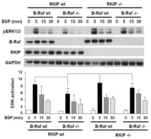
Figure 6. Depletion of RKIP rescues decreased MAPK signaling in B-Raf knockout primary keratinocytes.
Primary keratinocytes isolated form wild type (WT) and RKIP−/− and/or B-Raf−/− mice were serum-starved overnight prior to stimulation with EGF (5ng/ml) for different times as indicated. B-Raf, RKIP, pERK1/2 and GAPDH (loading control) in total cell lysates were detected by western blotting. The bar charts represent the densitometric quantification of pERK/GAPDH ratio in untreated (0) or EGF-treated keratinocytes from three independent experiments. Error bars represent S.D. *p<0.01.

