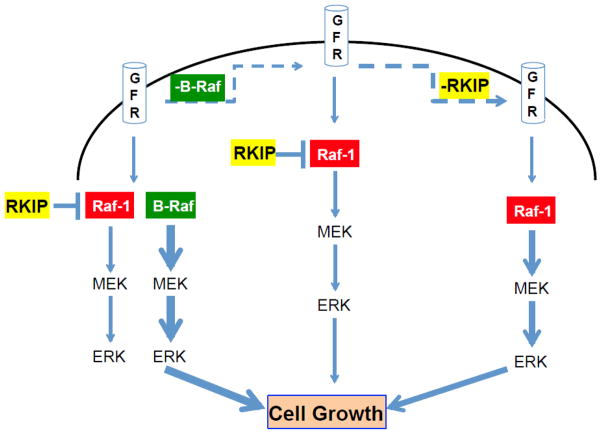
Figure 8. Schematic depicting RKIP regulation of MAPK pathway in MEFs.
(A) In normal conditions, activation of growth factor receptors (GFR) lead to activation of the MAPK (ERK) pathway mainly through B-Raf. In this condition Raf-1 is repressed by RKIP and only marginally participates in the signaling. (B) When B-Raf is depleted or deficient, signaling through MAPK is severely hindered since RKIP prevents full activation of Raf-1 and downstream phosphorylation of ERK. (C) When RKIP and B-Raf are deficient or depleted, signaling to ERK occurs through Raf-1. The thickness of the blue arrows is proportional to the intensity of the signaling.

