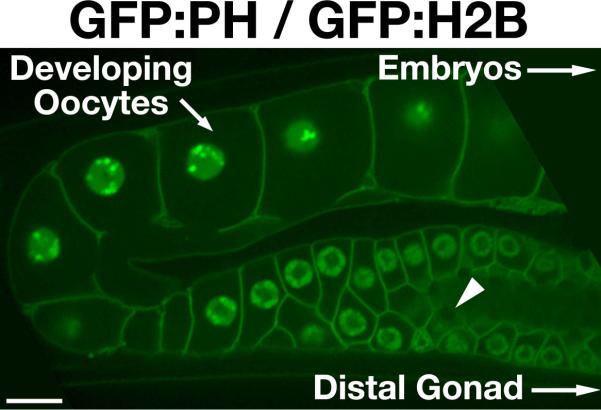
Figure 1.

Organization of the C. elegans syncytial gonad. Swept field confocal optics were used to image a transgenic animal co-expressing GFP fusions to histone H2B (HIS-58), to mark the DNA, and the PH domain of rat PLC1δ, which localizes to the plasma membrane. The gonad is a syncytial tube that contains multiple nuclei at various cell cycle stages. In the distal region (not shown), mitotic nuclei continually proliferate, feeding additional germ cells into the system. As the nuclei progress and mature, they enter different stages of meiotic prophase and produce mRNA. Translated messages generate protein that is loaded into developing oocytes through structures resembling ring canals (example highlighted by an arrowhead). Only the final 4–5 oocytes are diffusionally restricted from the syncytium. As oocytes pass through the spermatheca, they are fertilized and resume cell cycle progression. Ovulation is highly stereotypic, occurring every 22–24 minutes in fertile animals. Introduction of dsRNA triggers degradation of target mRNA in the gonad. Remaining protein present at the time of dsRNA delivery is depleted by continual packaging of gonad cytoplasm into developing oocytes. Within 36–48 hours following dsRNA treatment, the majority of target protein is depleted (typically >95% following injection of dsRNA), and the first embryonic cell division can be analyzed. Scale bar, 10 μm.
