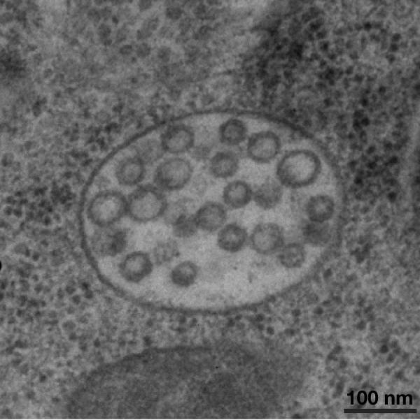
Figure 3.

Morphology of multivesicular endosomes that form within the C. elegans one-cell stage embryo. Intact animals were subject to high pressure freezing, followed by freeze substitution and embedding in plastic. Thin sections (60 nm) were cut using an ultramicrotome, and processed for imaging using a Philips CM120 electron microscope. Embryos at the one cell stage contain numerous multivesicular endosomes that range in size from 400–500 nm in diameter. The majority of intralumenal vesicles observed were 47–50 nm in diameter. The stereotypic ovulation of oocytes in C. elegans, which delivers newly fertilized embryos into the uterus directly adjacent to the spermatheca every 22–24 minutes provides the spatial cues necessary to study multivesicular endosome dynamics following high pressure freezing and EM analysis of intact animals. Artificial stimulation of receptor downregulation, a commonly used technique in other metazoan systems to analyze requirements for protein transport to the lysosome, is unnecessary in C. elegans. Oocyte maturation and fertilization reproducibly trigger the internalization and degradation of multiple transmembrane cargoes in one-cell stage embryos. Scale bar, 100 nm.
