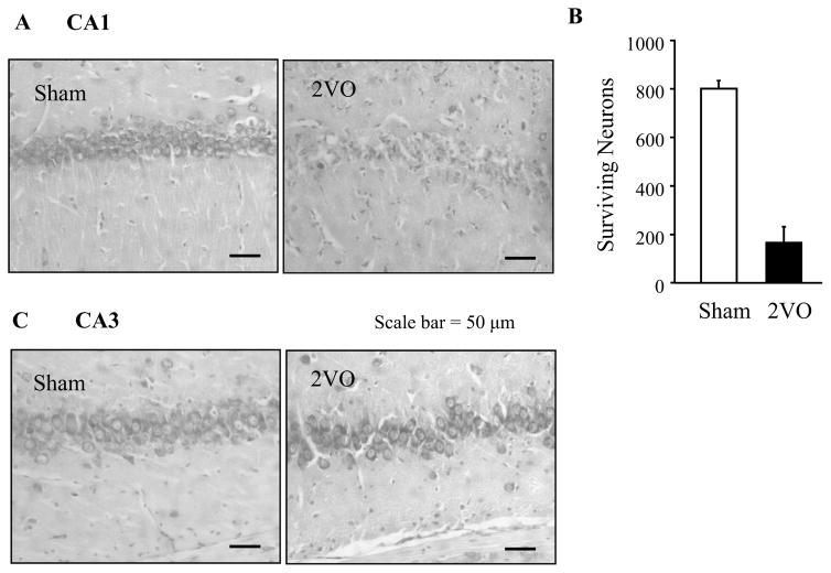
Fig 3.
Surviving CA1 hippocampal cells 4 days after the bilateral occlusion of common carotid arteries (2VO). (A) Photomicrograph showing a representative CA1 region from a sham and 2VO-treated rat. (B) Surviving Pyramidal cells per millimeter in the dorsal hippocampal CA1 region were counted in four 10 μm sections 120 μm apart, from and caudal to −4.2 mm relative to the bregma. The four values were summed for each rat. Data for each group of rats were expressed as the mean±SD (n=6). (C) A representative CA3 region from a sham and 2VO-treated rat.

