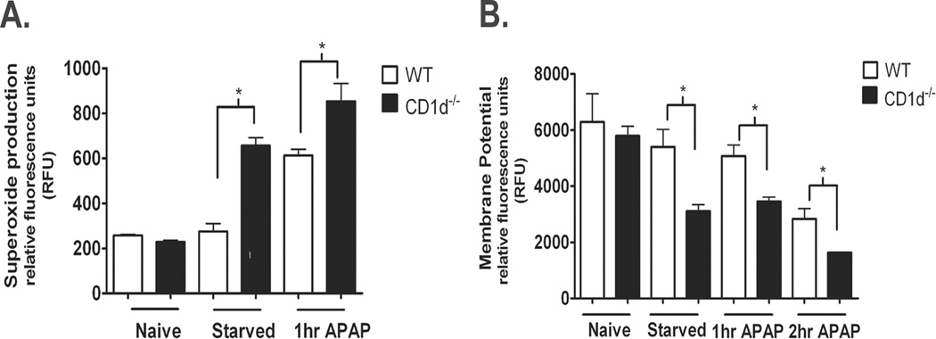
Figure 3. Increased mitochondrial dysfunction and ROS generation in CD1d−/− mice compared to WT after starvation and APAP-challenge.

Female WT and CD1d−/− mice were starved overnight for 16 h prior to i.p. injected with APAP (350 mg/kg). After 0, 1, or 2 h of APAP treatment, mice were sacrificed and mitochondria isolated. A) Mitochondrial polarization was detected by fluorometric analysis using JC-1 cationic dye. B) Mitochondrial ROS was detected by using MitoSOX. Results represent mean ± SEM of 3 mice per group. * p < 0.05 versus WT mice. Data shown are representative of two independent experiments.
