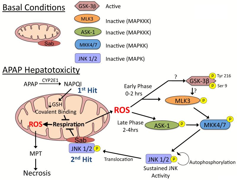
Figure 3. JNK activation during APAP-induced liver injury.
A. Under basal conditions most kinases, GSK-3β being a noted exception, are inactive and reside in the cytoplasm. B. APAP hepatotoxicity involves two hits to mitochondria. Mitochondrial GSH depletion and covalent binding by NAPQI (upstream hit to mitochondria) enhance mitochondrial ROS production. ROS cause two phases of MAPK activation during APAP hepatotoxicity, an early (0-2 hours) and a late (2-4 hours) phase, which involve different initiation events. The early phase likely involves GSK-3β activating MLK3, while the late phase is mediated by ASK-1 in the liver. Both then activate MKK 4/7, which in turn activates JNK. Once activated, JNK translocates to mitochondria and binds to Sab, a scaffold protein on the outer membrane of mitochondria. JNK binding to Sab leads to sustained enhancement of mitochondrial ROS generation which has two consequences: 1) self-amplifying activation of MAPK pathways and 2) MPT and collapse of mitochondrial function. MAPK = mitogen activated protein kinase; MAPKK = MAP kinase kinase; MAPKKK – MAPK kinase kinase kinase.

