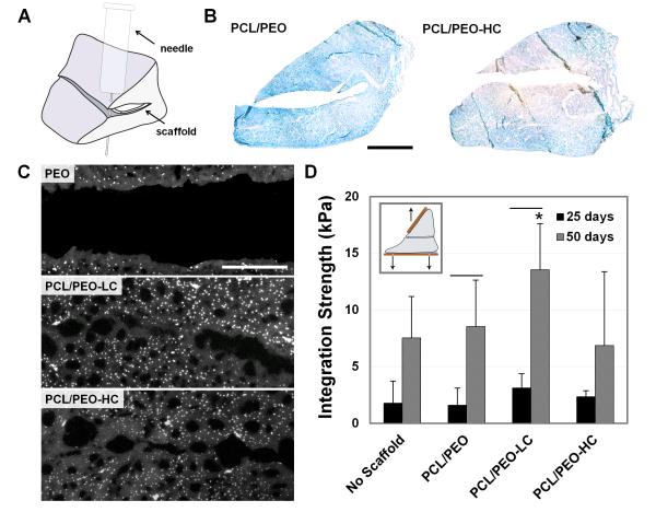
Figure 6.
Integrative repair of juvenile meniscus treated with collagenase-releasing scaffolds. (A) Schematic of repair construct with scaffold placed inside a horizontal meniscal tear. (B) AB staining of meniscus on day 7 with insertion of control and of collagenase-releasing composite scaffolds (n=3). Scale = 5 mm. (C) DAPI staining of wound interface after 50 days of culture with PCL/PEO, PCL/PEO-LC, and PCL/PEO-HC scaffolds (n=2). Scale = 0.25 mm. (D) Integration strength as a function of culture duration. Inset shows mechanical testing setup (n=5-6). * = p≤0.05 compared to PCL/PEO-HC. Line = p≤0.05 between 25 and 50 days.

