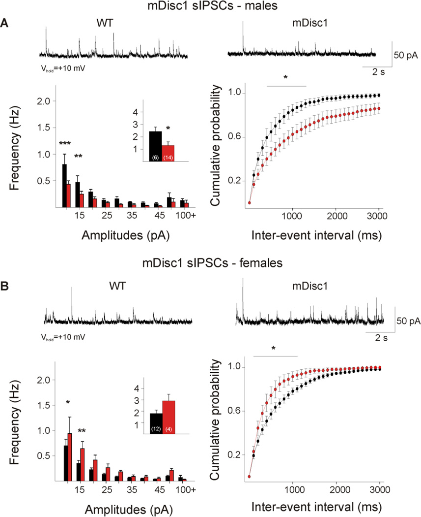
Figure 2. Sex differences in mDisc1 GABAergic synaptic activity.
A. Typical traces of sIPSCs recorded at +10 mV from male mDisc1 cortical pyramidal neurons. Note the significant decrease in mean sIPSC frequency (inset). Amplitude-frequency histograms indicated a significant decrease in small-amplitude events at the 10 and 15 pA bins. A significant difference was observed in the cumulative distributions of IEIs from 500–1400 ms. B. Typical traces of sIPSCs recorded at +10 mV from female mDisc1 cortical pyramidal neurons, showing a trend towards an increase in mean sIPSC frequency (inset). Note the significant increase in events at the 10 and 15 pA bins. A significant difference occurred in the cumulative distributions of IEIs from 200–1200 ms intervals.

