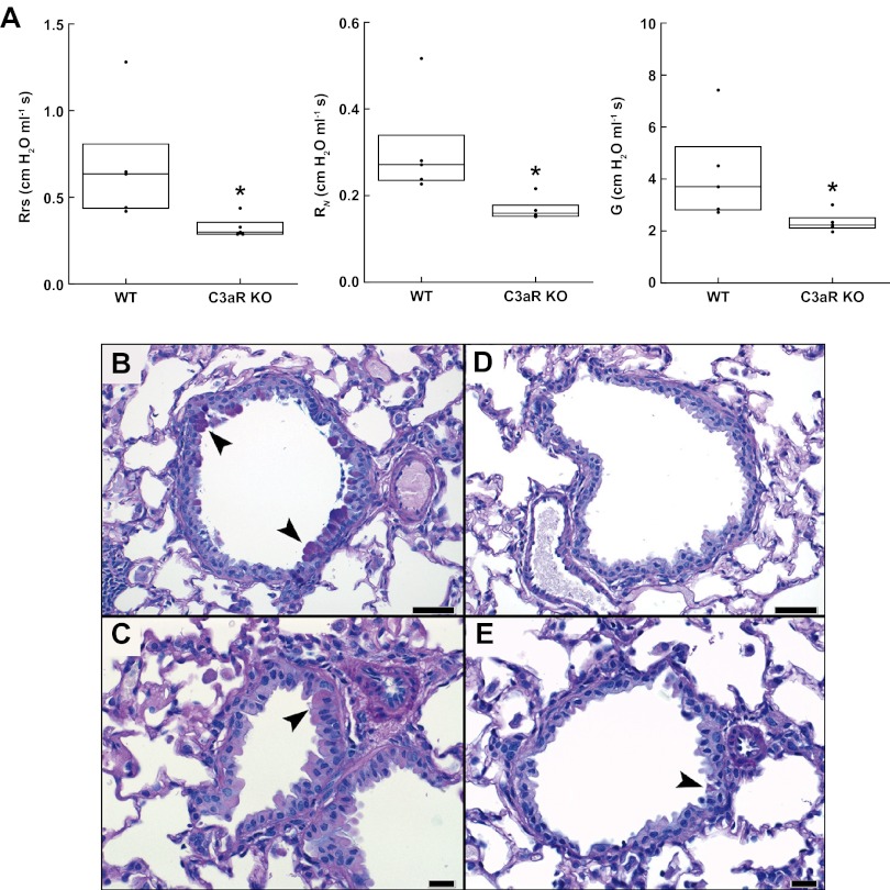
FIG 3 .
Physiological and histological analysis of airway inflammation in wild-type and complement-deficient mice. (A) Comparison of respiratory system resistance (Rrs), Newtonian resistance (RN), and tissue damping (G) in WT and C3aRKO mice after sensitization with Aspergillus antigens and chitin. Box plots indicate medians and interquartile ranges for each group. *, P = 0.016 (Rrs), 0.009 (RN), and 0.028 (G) compared with the WT group. (B) Photomicrographs of alcian blue PAS-stained, formalin-fixed, paraffin-embedded lung sections from WT (B, C) and C3aR KO (D, E) mice. Medium-caliber airways (above), bar = 50 µm; and smaller-caliber nonrespiratory airways (below), bar = 20 µm. The arrowheads depict epithelial cells with apical intracytoplasmic granules exhibiting positive PAS staining (goblet cells). Images are representative of an average of seven sized-matched large airways and eight size-matched small airways.

