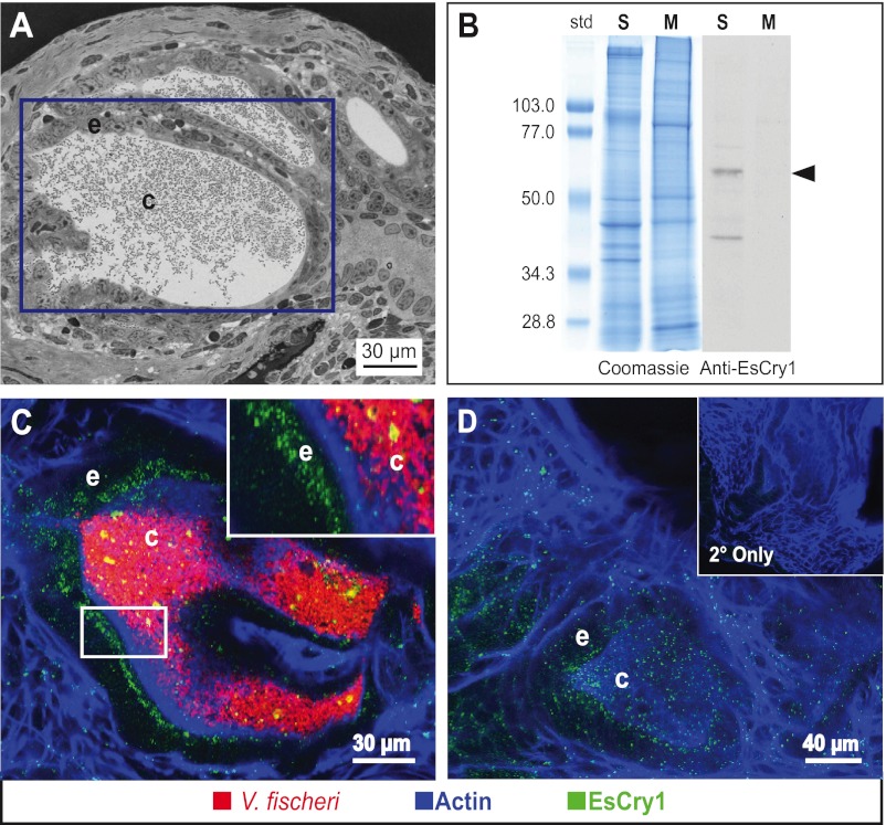
FIG 3 .
EsCry1 protein production in the light organ. (A) Light micrograph of a cross section of the E. scolopes light organ, shown in Fig. 1B. The purple box denotes the placement of crypt 1, which is comprised of an epithelial cell layer (e) surrounding a population of V. fischeri bacteria in the crypt lumen (c). (B) Western blot showing the immunoreactivity of the anti-EsCry1 antibody. Both aqueous soluble (S) and membrane (M) protein extracts from whole squid were run on SDS-PAGE gels and either stained with Coomassie blue (Coomassie) or transferred to a membrane and exposed to an anti-EsCry1 antibody (Anti-EsCry1). An arrowhead shows a major band at the predicted molecular mass of 62.3 kDa. Standards to the left (std) are shown in kDa. (C) Confocal micrograph of a colonized light organ stained with the anti-EsCry1 antibody. (D) Confocal micrograph showing an uncolonized light-organ crypt stained with the anti-EsCry1 antibody. The inset contains a negative control where the light organ was stained only with a secondary antibody (2° Only). In panels C and D, anti-EsCry1 is in green, V. fischeri cells are in red, and filamentous actin is blue. e, crypt epithelium; c, crypt lumen.

