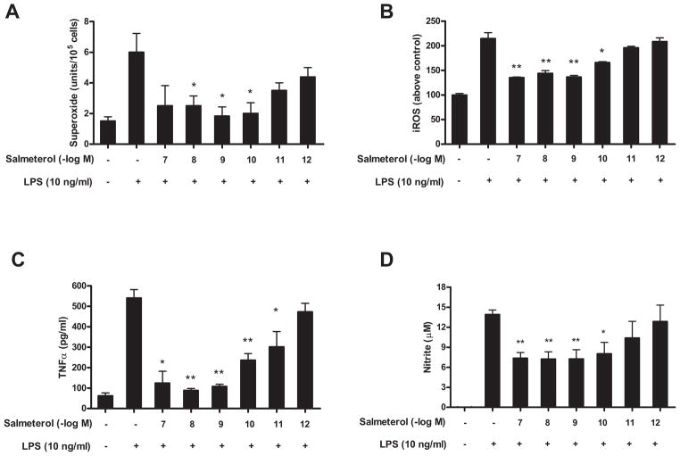
FIGURE 6.
Effect of salmeterol on LPS-induced production of pro-inflammatory factors from microglia. Microglia-enriched cultures were seeded at a density of 1×10⁵/well. Cells were pretreated with vehicle or various concentrations of salmeterol for 30 min followed by the addition of LPS. The production of LPS-induced extracellular superoxide production (A) was measured as SOD-inhabitable reduction of WST-1. LPS-induced intracellular ROS was determined by probe DCFH-DA (B). Salmererol’s effect on LPS-induced production of TNFα and nitrite were shown in Fig. 6C and Fig. 6D. Results were expressed as mean ± SE from three to five independent experiments in triplicate. *P<0.05, **P<0.01 compared with the LPS-treated cultures.

