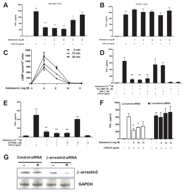
FIGURE 7.
Low dose salmeterol-mediated anti-inflammatory effects are β2AR/β-arrestin2 dependent, but cAMP/PKA and cAMP/EPAC-independent. Microglia culture prepared from C57/BL6 (A) or β2AR deficient mice (B) were pretreated with vehicle or indicated concentrations of salmeterol for 30 min prior to the addition of LPS. Supernatant were collected at 3h after LPS addition for TNFα analysis (A–B). Figure C: Enriched primary microglia were incubated with vehicle or indicated concentrations of salmeterol at 37°C for 5, 10, or 30 min. After incubation, the cells were lysed and cAMP levels were determined using a cAMP assay kit. Data were expressed as pmol cAMP per 1 million cells (C). Figures D and E: Enriched primary microglia cells were pretreated with vehicle or PKA inhibitors, including H89 (1 μM for 45 min), Rp-cAMP (50 μM for 45 min) (D), or EPAC agonist 8CPT-2′-O-Me-cAMP (CPTOMe) (10 μM for 45 min) (E) prior to stimulation with salmeterol (10−10M) and LPS (5 ng/ml). Supernatants were collected 3h after LPS addition to measure TNFα levels. Figure F and G: Primary microglia cells were transfected with 100 pmol specific β-arrestin2 siRNA or control siRNA, and 48 hrs after transfection cells were treated with indicated concentrations of salmeterol for 30 min prior to addition of LPS. Supernatants were collected for TNFα assay (F). Knockdown of expression of the β-arrestin2 was determined by western blot analysis (G). Results in A–F were expressed as mean ± SE from three to four independent experiments in triplicate. *P<0.05, **P<0.01 compared with the LPS-treated cultures.

