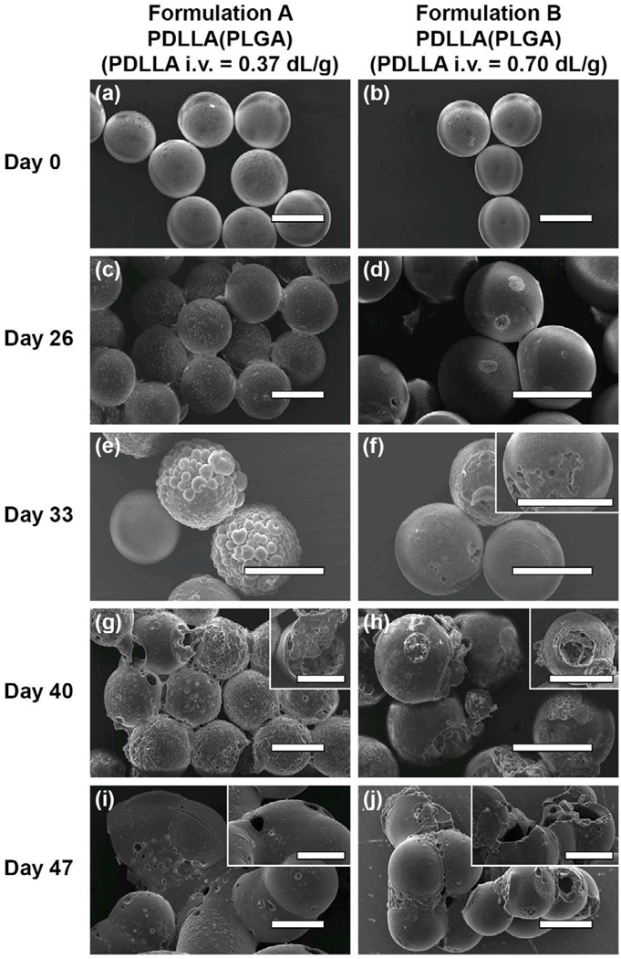
Figure 3.
SEM images depicting the surface morphology of double-walled PDLLA(PLGA) microspheres with a low PDLLA molecular weight shell layer (formulation A) and a high PDLLA molecular weight shell layer (formulation B) at different stages of the degradation process. (a) and (b) are images of initial microspheres before degradation, (c) and (d) 26 days, (e) and (f) 33 days, (g) and (h) 40 days, and (i) and (j) 47 days after degradation. The inserts show microspheres with pore or cavity formation. Scale bar = 50 µm.

