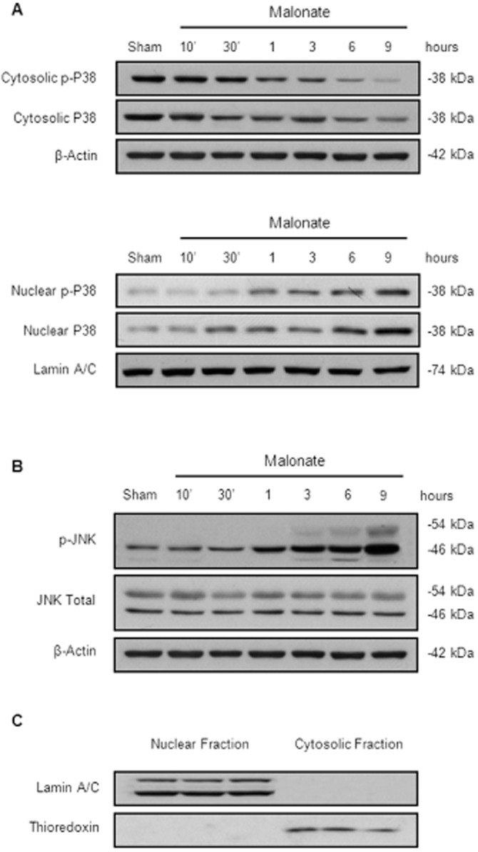
Figure 3.

Time course activation of SAPK induced by malonate. Animals were killed at different time points after intrastriatal injection of malonate (1.5 μmol/2 μL). (A) Malonate induces p38 translocation to the nucleus in a time-dependent manner. Representative Western blots of cytosolic and nuclear fractions. β-actin and lamin A/C were used as equal loading control (n = 5 per group). (B) Representative Western blots showing the time course expression levels of p-JNK in the cytosol after malonate administration (n = 5 per group). (C) Representative blots showing the purity of our samples.
