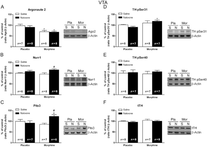
Figure 6.
Nurr1 and Pitx3 expression is activated in the VTA from morphine-withdrawn rats. Representative Western blots and semi-quantitative analysis of Argonaute 2 (A), Nurr1 (B), Pitx3 (C), pSer31-TH (D), pSer40-TH (E) and total TH (tTH; F) protein levels in the VTA tissue isolated from placebo (pla) or morphine (mor)-dependent rats after s.c. administration of saline (s) or naloxone (n). Each bar represents the mean optical density ± SEM; values are expressed as % of controls. Newman–Keuls' post hoc comparison test revealed a significant increase in Nurr1, Pitx3 and pSer31-TH in morphine-withdrawn rats at 2 h after naloxone injection, whereas there was a decrease in Argonaute 2 protein levels in morphine-dependent rats receiving saline and after naloxone-induced morphine withdrawal. No significant modifications were observed in the tTH protein levels #P < 0.05 versus morphine + saline; +P < 0.05 versus placebo + naloxone; *P < 0.05 versus placebo + saline.

