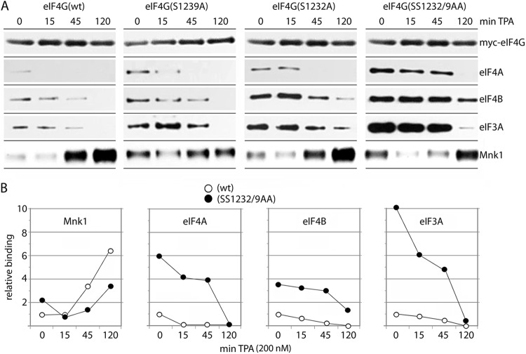
Fig 5.
Time course of TPA-induced binding of eIF4A, eIF4B, eIF3A, and Mnk1 with wt and mutant 1222 eIF4G fragments. HEK293 cells were transfected with the indicated 1222 fragments (16 h), serum starved (24 h), and treated with 200 nM TPA for the intervals shown. Lysates were subjected to IP/immunoblotting with the indicated antibodies. The experiments were repeated three times, and the results of a representative series are shown. (A) TPA-induced binding of eIF4A, -4B, and -3A to wt 1222, 1222(S1239A), 1222(S1232A), or 1222(SS1239/2AA). Due to very weak binding of the 1222 fragment with eIF4A (Fig. 1D and E), we exposed filters for extended intervals for detection of signal. (B) Quantitative chemiluminescence detection of the immunoblot signals for wt and double mutant 1222 fragments. Average values of the relative binding are shown; standard errors were <12% from the average values for 0 and 15 min of TPA stimulation and <15% for the 45- and 120-min time points.

