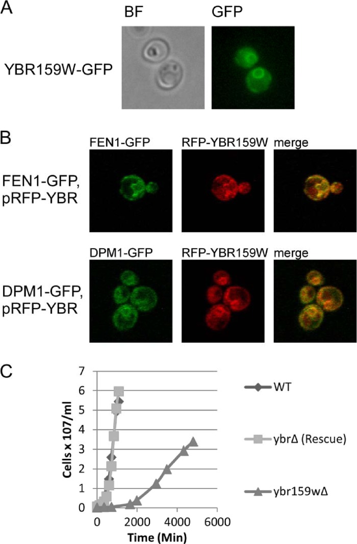
Fig 3.
Cellular analysis of YBR159W. (A) Live cell epifluorescence imaging of endogenously tagged YBR159W-GFP indicates YBR159W localizes mainly to the ER membrane. (B) Live cell confocal microscopy showing the colocalization of YBR159W with the VLCFA pathway enzyme FEN1 and ER membrane protein DPM1. YBR159W is expressed on a low-copy-number plasmid and tagged with DsRed. FEN1 and DPM1 are endogenously expressed and tagged with GFP. (C) Deletion of YBR159W results in a very slow growth rate.

