
Fig 1.
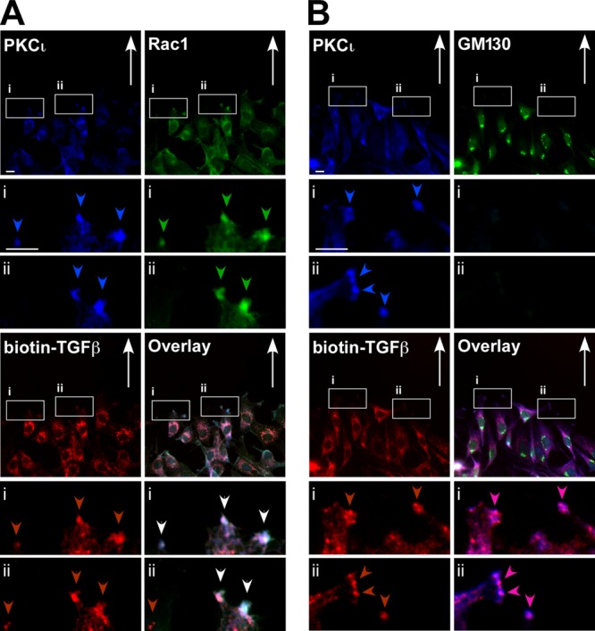
Atypical PKCι colocalizes with TGF-β receptors at the leading edge of migrating cells. Rat2 fibroblasts were scratched and incubated for 4 h to establish cell polarity and then fixed, permeabilized, and immunostained with anti-PKCι (PKCι, blue), biotin-labeled TGF-β (biotin-TGF-β; red), anti-Rac1 (Rac1, green [A]), or anti-GM130 (Golgi apparatus marker, green [B]). The direction of cell migration is indicated with arrows, and PKCι, Rac1, and TGF-β receptors at the leading edge of migrating cells are indicated by blue, green, and red arrowheads, respectively. The white arrowheads indicate the colocalization of all three proteins. Bar, 10 μm.