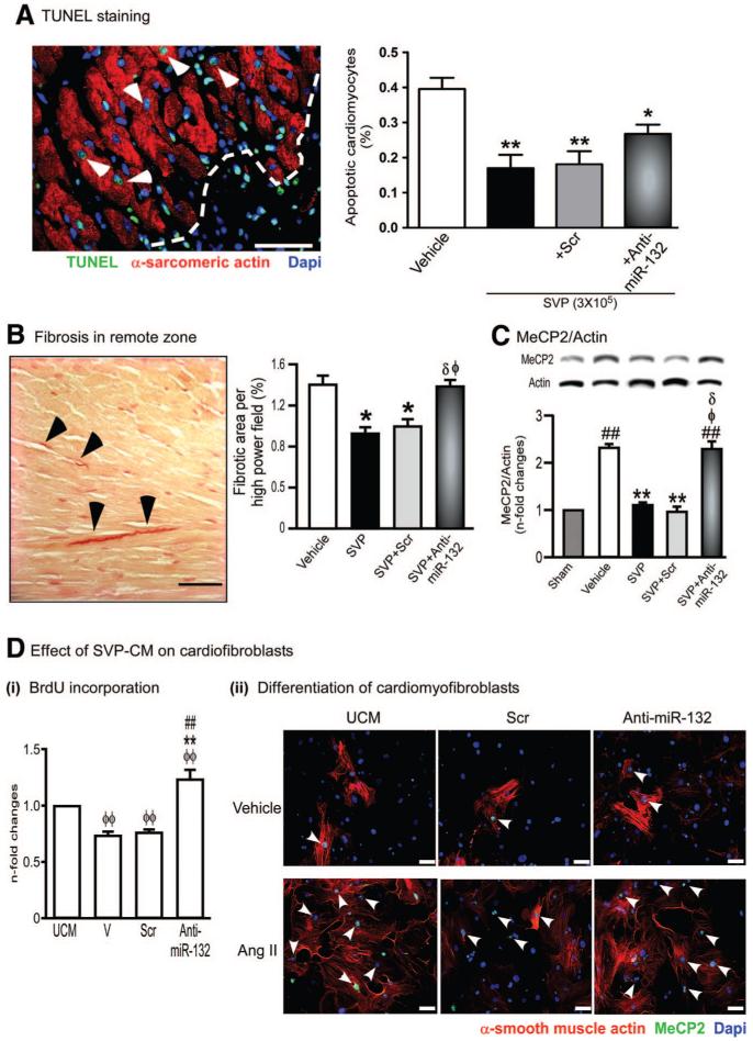
Figure 7. SVP transplantation attenuates cardiomyocyte apoptosis and interstitial fibrosis.
A, Representative immunofluorescence image and bar graph showing abundance of apoptotic cardiomyocytes at 14 days post-MI in hearts injected with vehicle or transplanted with naïve, scrambled-transfected or antimiR-132-transfected SVPs. Data are means±SE (n=5 mice per group). *P<0.05 and **P<0.01 vs vehicle. B, Representative image and bar graph showing fibrosis in the spared myocardium. C, Representative immunoblots and bar graph showing the expression of MeCP2 in myocardium (n=5 mice per group). ##P<0.01 vs Sham; **P<0.01 vs Vehicle; δP<0.05 vs nontransfected SVPs; ɸP<0.05 vs SCrtransfected SVPs. D, Bar graph (i) and immunocytochemical images (ii) showing the effect of SVP-CM on BrdU incorporation (i) and differentiation of murine cardiac fibroblasts into myofibroblasts (ii). Cardiac fibroblasts were treated with Angiotensin II (Ang II, 100 nmol/L) for 24 hours to induce differentiation into myofibroblasts in the presence or absence of SVP-CM. Differentiated myofibroblasts are identified by positive staining for α-smooth muscle actin and MeCP2 (arrow head). Data are means±SE of experiments performed in quadruplicate. ɸɸP<0.01 vs unconditioned medium (UCM); **P<0.01 vs vehicle (V); ##P<0.01 vs scrambled sequence (Scr). Scale bars are 50 μm.

