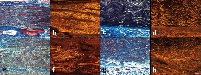
Figure 2.

Injury zones at five days (a–d, bar = 200 µm) and 65 days (e–h, bar = 50 µm), comparing crush (top) to experimental (bottom) injuries; Masson's trichrome and neurofilament. Note the aberrant axonal sprouting and regeneration in the experimental injury group, associated with increased intrafascicular collagen, in contrast to orderly regeneration and lack of scar in the simple crush group.
