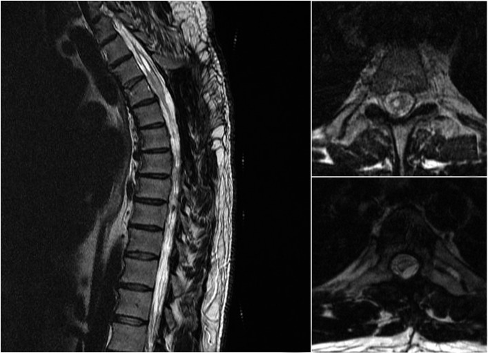
Figure 4.
Postoperative T2-weighted MRI scan a mid-sagittal view demonstrates interval decompression of the spinal cord at the T4–5 level. The persistent syrinx has decreased substantially in size b axial view at the level of T3 showing interval decrease in syrinx size c axial view at the T4 level showing re-expansion of the thoracic cord following laminectomy and resection of calcified arachnoid

