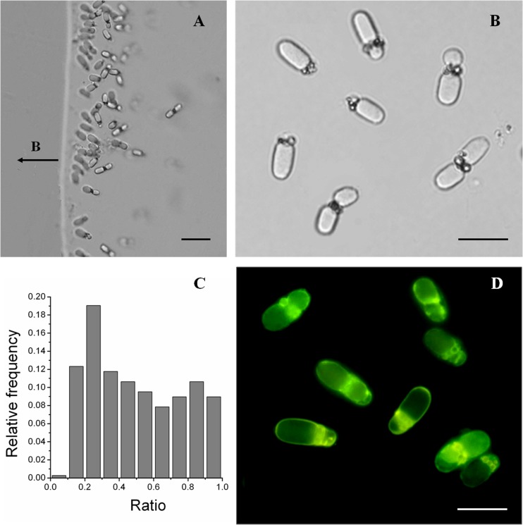
Fig 1.
Morphology of large, rod-shaped MTB based on optical microscopy. DIC images of large, rod-shaped MTB are shown in panels A and B. The ratio distribution of the lengths of the two parts is shown in panel C. Panel D shows the fluorescence of large, rod-shaped MTB exposed to blue light (wavelength, 450 to 480 nm). Scale bars, 20 μm in panel A and 10 μm in panels B and D.

