
Fig 3.
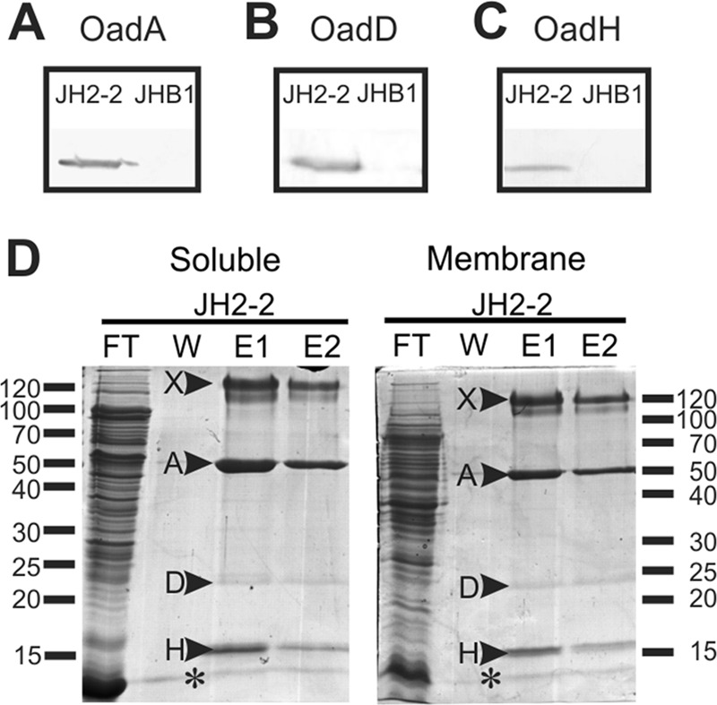
Isolation and identification of Ef-OAD subunits. Western blotting for the detection of Ef-A (A), Ef-D (B), and Ef-H (C) in crude extracts of wild-type (JH2-2) and citO mutant (JHB1) strains. To detect the presence of Ef-A and Ef-H subunits, polyclonal antibodies prepared in the laboratory were used (see Materials and Methods), whereas for the Ef-D protein, alkaline phosphatase-conjugated streptavidin, which permits the detection of biotinylated proteins, was utilized. (D) Soluble and membrane protein extracts corresponding to the wild type (JH2-2) were passed through an avidin resin. After washing (W), total biotinylated proteins were eluted with buffer containing 5 mM avidin. Two elution fractions (E1 and E2) were recovered, and all fractions were then run in an SDS-PAGE gel. Indicated bands (arrowheads) were excised from the gel and identified by mass spectrometry. Band A, Ef-A; band D, Ef-D; band H, Ef-H; band X, Ef-D protein aggregate. FT, flowthrough. An asterisk corresponds to avidin released from the column.