Fig 4.
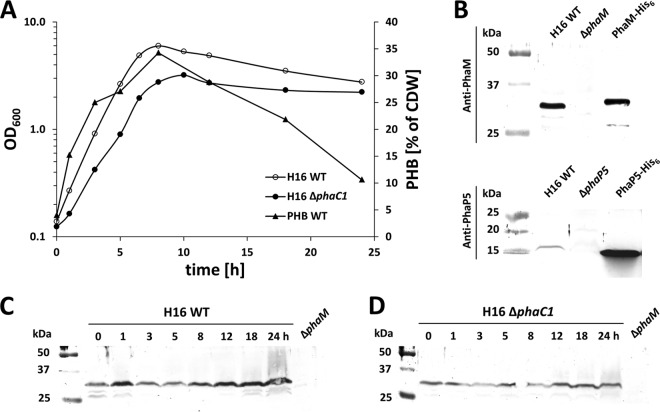
Expression of PhaM and PhaP5 and accumulation of PHB. R. eutropha H16 and the R. eutropha ΔphaC1 strain were grown on NB-gluconate medium, and optical densities at 600 nm and PHB contents of samples were recorded (A). CDW, dry weight of cells. Antisera against PhaM or PhaP5 reacted with respective proteins of the expected size in R. eutropha and with purified PhaM-His6 or PhaP5-His6, whereas no signals were observed in the respective deletion strains (H16 ΔphaM or ΔphaP5) (B). Expression of PhaM in H16 (C) and H16 ΔphaC1 (D) at different stages of growth was determined in whole-cell extracts by Western blotting (immunoblotting). Comparable loading of wells was performed using the same OD600 equivalents and checked by SDS-PAGE and Coomassie brilliant blue staining (not shown). Purified PhaM-His6 or PhaP5-His6 was used as a positive control, and whole-cell extracts of H16 ΔphaM and H16 ΔphaP5 corresponding to the 0-h time point were used as negative controls. One growth experiment with the respective Western blot out of two biological replicates is shown. WT, wild type.

