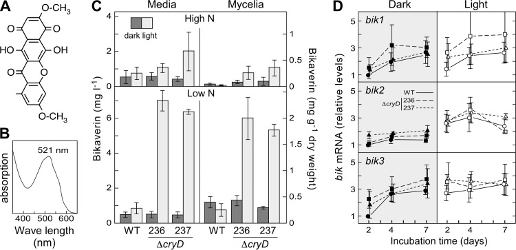
Fig 7.
Effect of the ΔcryD mutation on bikaverin production. (A) Chemical structure of bikaverin. (B) Representative example of absorption spectrum of the red pigment isolated from the shake cultures of the ΔcryD mutants SF236 and SF237 grown in low-N medium in the light (Fig. 4C). (C) Bikaverin concentrations in the mycelia and the filtrates of shake cultures of the same strains grown for 7 days at 30°C in high-N or low-N media in the dark or under constant illumination (3 W m−2). (D) qRT-PCR analyses for genes bik1, bik2, and bik3 in RNA samples from the mycelia of cultures after 2, 4, and 7 days of incubation in low-N medium. Relative expression was referred to the value of the wild type in the dark. The data on bikaverin and bik mRNA levels show averages and standard errors from three independent experiments.

