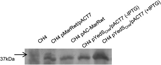
Fig 2.

Urea SDS-PAGE analysis of outer membrane proteins. Outer membrane proteins were purified and subjected to gel electrophoresis as described previously (27). The arrow to the left denotes the migration of the 37-kDa molecular weight marker. Each lane was loaded with 5 μg of total outer membrane protein.
