
Fig 1.
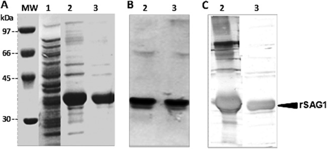
Expression and purification analysis. SDS-PAGE analysis of bacterial lysis extract and Coomassie blue staining of rSAG1 antigen (A) and Western blot treated with antipolyhistidine-peroxidase conjugate antibody (B) or a human reference serum sample (C). Numbers correspond to the noninduced cell culture (lane 1), the crude solubilized preparation with 8 M guanidine-HCl (lanes 2), and the purified solubilized preparation under denaturing conditions (6 M urea) (lanes 3). Molecular mass (MW) markers in kDa are indicated on the left.